| Homepage | Hand | Wrist | Forearm | Elbow | Knee | Ankle | Foot |
The Appendicular Skeleton
Anatomy

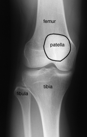
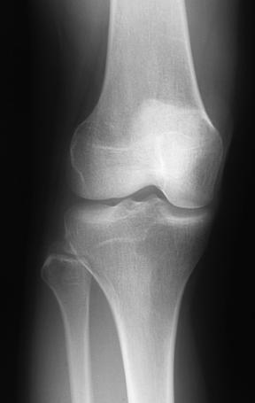
AP Knee
The knee is a synovial joint. The medial and lateral condyles of the femur, the patella, and the medial and lateral condyles of the tibia make up the knee joint. The lateral condyle of the femur is slightly larger than the medial condyle as it supports more of the body weight.
The patella is the largest sesamoid bone in the body and lies in the quadriceps femoris muscle on the anterior aspect of the knee joint. The medial and lateral condyles of the tibia supply the weight-bearing surfaces of the knee joint. The fibula does not form part of the knee joint as it is not constructed to be weight-bearing.

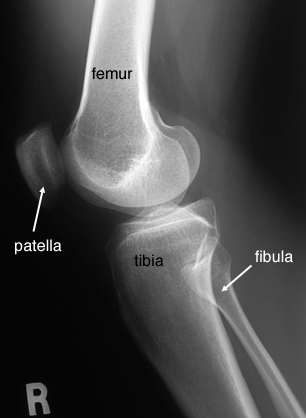
Lateral Knee
The medial and lateral condyles of the femur articulate with those of the tibia. The patella articulates with the patellar articular area between the femoral condyles.
